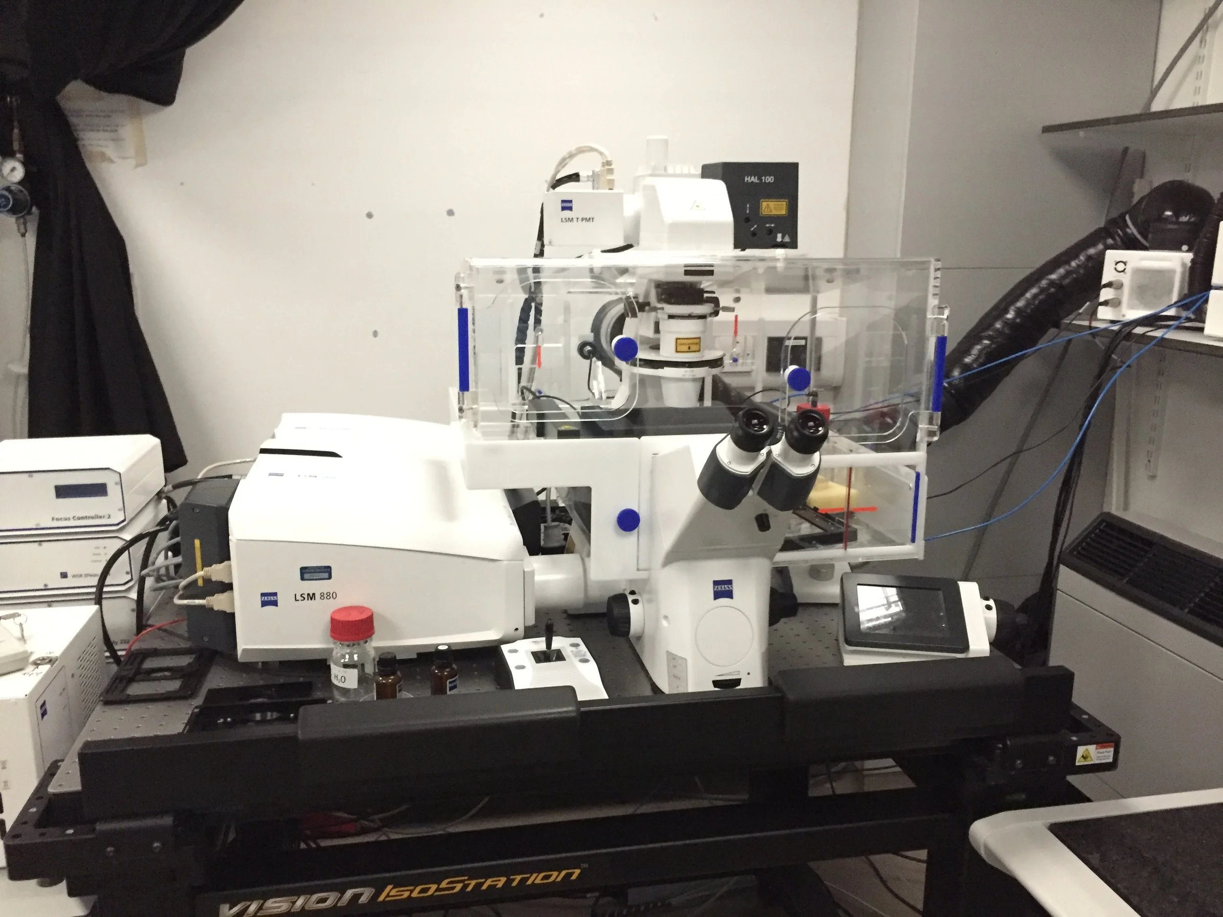
We need to look at tiny cells and structures that can’t be seen with the human eye to do our research at the Gurdon Institute. New microscopes and techniques are being developed all the time and we use specially designed microscopes to take different images. You’ll find some examples below!
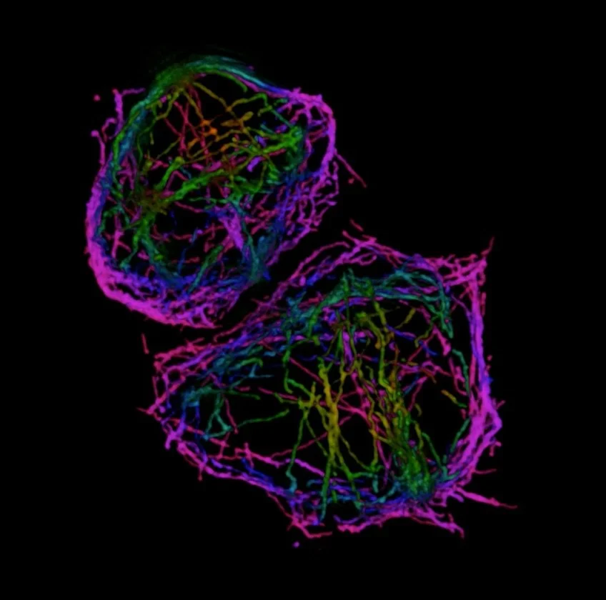
The internal structure of the protective blood cells (macrophages) in a fruit fly by Dr Nicola Lawrence.
This was taken using a Structured Illumination Microscope.
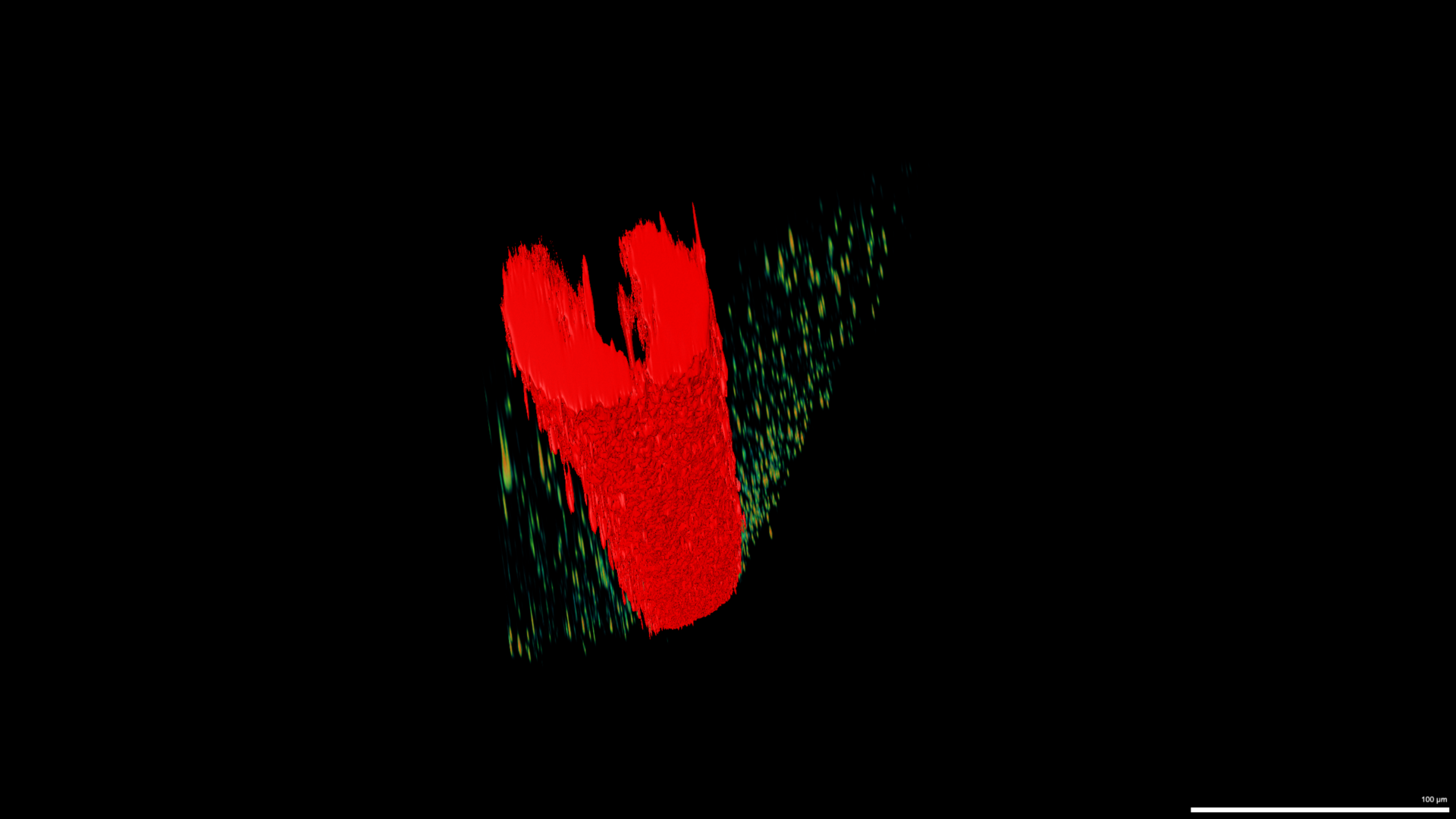
3D image of a chicken embryo’s neural tube - this will later become the spine taken by Ren Moon on a Lightsheet Microscope.
Image of the egg chamber in an adult female Fruit Fly by Dmitry Nashchekin.


