
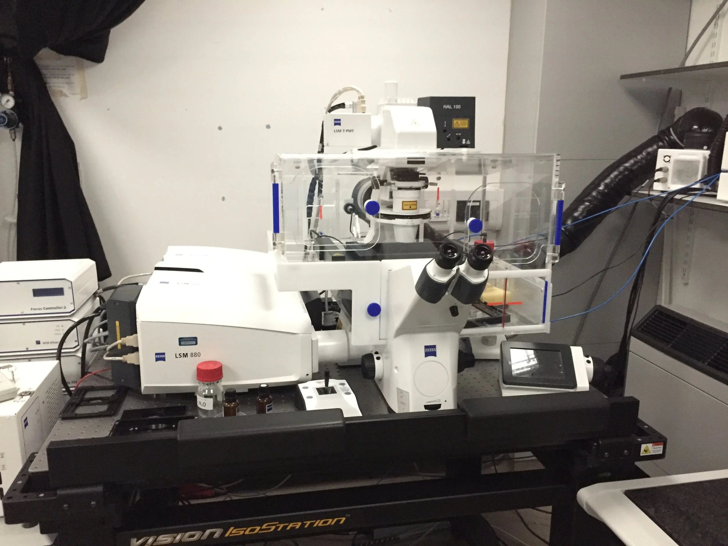
We need to look at tiny cells and structures that can’t be seen with the human eye to do our research at the Gurdon Institute. New microscopes and techniques are being developed all the time and we use specially designed microscopes to get different kinds of information from our samples. You’ll find some examples below!
The internal structure of the protective blood cells (macrophages) in a fruit fly larva prepared and imaged by Dr Nicola Lawrence. These are dome shaped cells; the yellow and green sections in the image are closer to you with the purple and pink sections, further away.
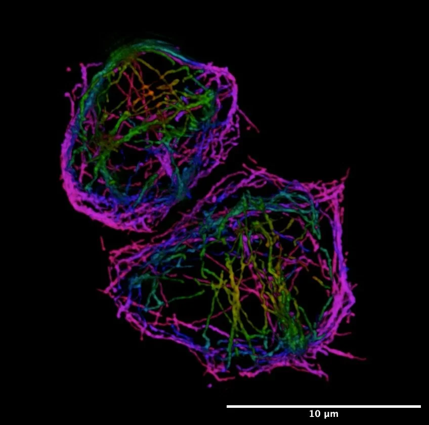
These labelled structures within the cells are less than a ten-thousandth of a mm across (!) and we need a Super-Resolution Microscope to see them.
3D image of a chicken embryo’s neural tube which will later become the spine taken by Ren Moon on a Lightsheet Microscope. The neural tube is a 3 dimensional structure and we need to use a microscope that can image in 3 dimensions to study it as it develops.

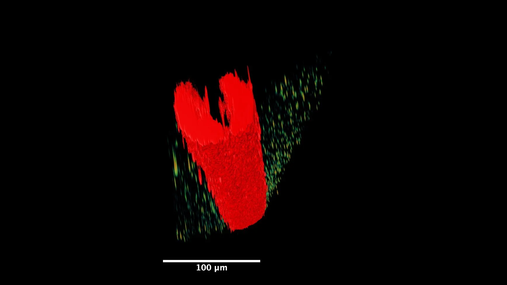
The egg chamber (a future egg cell is outlined in orange) in an adult female fruit fly prepared and imaged by Dr Dmitry Nashchekin. In this image, we were interested in how microtubules, special structures involved in transporting molecules around cells, work. The ends of the microtubules are shown here in green and red and we needed to create a time-lapse video using a spinning-disc confocal microscope to see how these structures move.